1.4 Neuroendocrine Pathway of Milk Secretion
Milk synthesis and release are controlled primarily by the effect of suckling on hormonal release via a complex neuroendocrine process. Two of the major hormones involved in this process are prolactin and oxytocin.
The sensory nerve endings of the nipple and the areola are stimulated by the infant’s suckling at the breast, which also stimulates prolactin release from the anterior pituitary. Prolactin and oxytocin are secreted and released into the circulation via a neural reflex pathway. Oxytocin may also be released by other sensory pathways ie. visual or auditory. Prolactin stimulates milk synthesis and secretion in the lactocytes. Under the influence of oxytocin, the myoepithelial cells enclosing the alveoli and terminal milk ducts contract and press the milk from the alveoli into the milk ducts (milk ejection). This is commonly known as the let-down reflex (Blackburn 2013).
The mammary glands adjust the milk supply according to infant demand. Frequent breastfeeding during the first days of lactation results in:
- frequent stimulation of the sensory nerve endings of the breast and
- complete removal of the milk from the breast
both of which result in increased milk supply over time.

Figure 5: Neuroendocrine Pathway of Milk Secretion
Source: ENeA own
Prolactin
 Prolactin stimulates the production of milk proteins and fat in the alveolar secretory cells in conj unction with the hormones insulin and cortisol. Prolactin levels increase markedly towards the end of a feed, peaking approximately 45 mins after the onset of suckling, in order to increase the volume, fat and protein content of the breast milk in preparation for the subsequent feed (Blackburn 2013). It also maintains milk production (galactopoiesis) by stimulating milk synthesis and secretion in the lactocytes.
Prolactin secretion is subject to a multifactorial control cycle which is regulated by a number of inhibiting factors:
- Prior to delivery prolactin levels are suppressed by the presence of high levels progesterone and estrogens, which inhibit prolactin secretion. Following delivery, progesterone and estrogen levels decrease facilitating a physiological response to prolactin.
- Secretion of prolactin is, by default, inhibited by the hypothalamus. As a result of nerve stimulation by suckling the secretion of prolactin-inhibiting factor (PIF) from the hypothalamus decreases, removing the inhibitory mechanism and resulting in increased prolactin secretion (Blackburn 2013).
Studies have shown that prolactin levels are highest during the early postpartum period and decline steadily by six months postpartum, however it appears this does not significantly affect breastfeeding frequency or milk production (Cox et al. 1996). Prolactin has also been shown to be strongly circadian dependent with highest levels measured at night when the rate of milk synthesis is also at its highest thereby resulting in higher prolactin levels in the first morning feed (Cregan et al. 2002).
Oxytocin
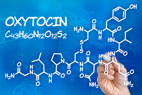 Oxytocin release during lactation is responsible for contraction of the myoepithelial cells in the mammary gland which forces milk from the alveoli into the milk ducts (milk ejection) (White-Traut et al. 2009).
Oxytocin is synthesized in the nucleoli supraopticus and paraventricularis of the hypothalamus and is stored in the posterior pituitary (White-Traut et al. 2009). It is released through stimulation of the breast by the suckling infant during lactation. Visual or auditory stimulation such as the crying of an infant or thinking about breastfeeding may also release oxytocin from the posterior pituitary (Blackburn 2013).
Additionally, oxytocin causes the myometrium of the uterus to contract. This allows the uterus to return to its pre-pregnancy size but sometimes causes uncomfortable contractions during breastfeeding which are referred to as afterpains.